
What happens to the hippocampus during uncal herniation?
DOI:
https://doi.org/10.37085/nsa.2025.9Keywords:
Brain, Humans, Hippocampus, Limbic lobe, Intracranial hypertensionAbstract
Introduction
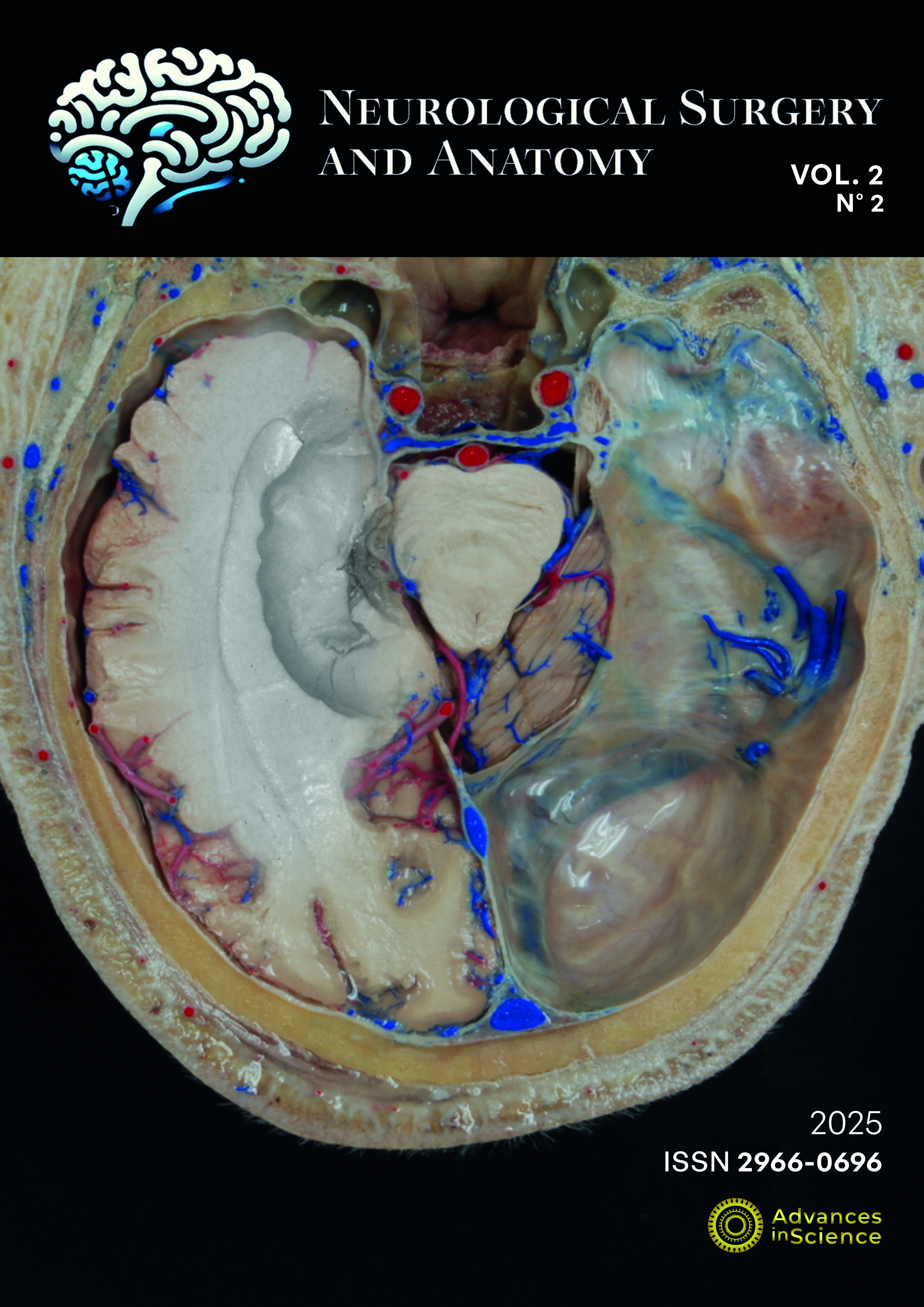
Uncal herniation is a well-known phenomenon linked to unchecked intracranial hypertension and associated with specific neurological syndromes. Its understanding is critical for medical students as it can the cause or be associated with death. The hippocampus, formed by cornus ammonis and dentate gyrus, is part of the limbic lobe, and its anatomical knowledge, a crucial part of the study of several diseases, including Alzheimer's.
Objective
To describe hippocampal anatomy using microsurgical anatomy images and Anatomage 10.0 data, evaluating the peri-mortem effects of uncal herniation.
Methods
Microsurgical dissection of formalin-fixed cadaveric-human brains exposed limits and parts of the hippocampal formation. Anatomage Table 10.0 is a technological tool for anatomy learning, which provides an interface for interaction with digitized data from human cadaveric donors. Both types of images were combined to allow identification of parts of hippocampal formation.
Results
Five cadaveric donors in Anatomage 10.0 showed unilateral or bilateral signs of uncal herniation. Amount of herniated tissue was inversely related to donor's age, suggesting age-related atrophy and/or different pathologies leading to death. Anatomage 10.0 allows layers of anatomical structures to be peeled back, highlighting the anatomical relationships under different degrees of uncal herniation.
Conclusions
The hippocampus is expected to be affected during uncal herniation, but this understanding is seldom reached by medical students exploring the limbic system. Anatomage 10.0 can expedite this realization, stimulating students to acquire more detailed anatomical terminology for adequately describing what they are seen. This contributes to deeper, clinically meaningful understanding of the brain.
References
Martins C. Rhoton’s Lab. World Neurosurg. 2016;92. doi: 10.1016/j.wneu.2016.06.035
Alasmari WA. Medical students’ feedback of applying the virtual dissection table (Anatomage) in learning anatomy: A cross-sectional descriptive study. Adv Med Educ Pract. 2021;12:1303–7. doi: 10.2147/AMEP.S324520
Ono M, Kubik S, Abernathey C. Atlas of Cerebral Sulci. NewYork: Thieme; 1990. 218.
Plum F, Posner J. Diagnostico de Estupor e Coma. 2nd ed. Rio de Janeiro: Guanabara Koogan; 1977. 360.
Rhoton Jr. A, Ono M. Microsurgical anatomy of the region of the tentorial incisura. In: Wilkins R, Rengachary SS, editors. Neurosurgery. 2nd ed. New York: McGraw Hill; 1996. p. 897–915.
Ono M, Rhoton AL, Barry M. Microsurgical anatomy of the region of the tentorial incisura. J Neurosurg. 1984;60:365–99.
Martins C, Felipe L, Tabarelli P, Campero A, Alencastro LF, Moraes Valença M, et al. Exploring the third ventricular floor anatomy: clinical insights for endoscopic third ventriculostomy. Neurological Surgery and Anatomy. 2024;2024(1):15–21. doi: 10.37085/ns&a.2024.5
Martins C, Quilis Quesada V, Cavalcanti A, Martins A, Moraes Valença M, Wen HT, et al. A handy tool to teach microneurosurgical anatomy of uncus. Neurological Surgery and Anatomy. 2024;2024(2):67–71. doi: 10.37085/ns&a.2024.15
Wijdicks E, Gary M. Transient locked-in syndrome after uncal herniation. Neurology. 1999;52(6):1296–7.
Katzir M, Attia M, Sviri GE, Zaaroor M. Uncal herniation in a fully conscious patient - The sliding uncus syndrome. Br J Neurosurg. 2015;29(2):308–9. doi: 10.3109/02688697.2014.977779
Udayakumaran S, Ben Sira L, Constantini S. Chronic uncal herniation secondary to posterior fossa shunting: Case report and literature review. Child’s Nervous System. 2010;26(2):267–71. doi: 10.1007/s00381-009-1027-z
Beucler N, Cungi P-J, Baucher G, Coze S, Dagain A, Roche P-H. The Kernohan-Woltman Notch Phenomenon : A Systematic Review of Clinical and Radiologic Presentation, Surgical Management, and Functional Prognosis. Review Article J Korean Neurosurg Soc. 2022;65(5):652–64. [accessed 13 Oct 2024] Available from: https://doi.org/10.3340/jkns.2022.0002
Duvernoy H., Cattin F., Risold P-Y. The Human Hippocampus. 4th Edition. Heidelberg: Springer-Verlag; 2013. 1–237 p.
Kier EL, Fulbright RK, Bronen RA. Limbic Lobe Embryology and Anatomy: Dissection and MR of the Medial Surface of the Fetal Cerebral Hemisphere. Vol. 16, AJNR Am J Neuroradiol. 1995.
Downloads
Published
Issue
Section
License
Copyright (c) 2025 Neurological Surgery and Anatomy

This work is licensed under a Creative Commons Attribution 4.0 International License.